


Finding a cure for Schimke Immuno Osseus Dysplasia (SIOD)
SIOD RESEARCH
We help to accelerate the development and delivery of new therapies and treatments, and facilitate a place of hope for SIOD families and their children with information and resources to help them navigate life with this disease.

Our lab does alot of work in conjunction with other doctors and labs. We are excited about the collaboration with Dr. Matthew Porteus, who is considered one of the pioneers and founders of the field of genome editing—a field that now encompasses thousands of labs and several new companies throughout the world.


NOVEMBER 2025 UPDATE:
A TIMELINE OF MILESTONES IN SIOD RESEARCH AND CLINICAL CARE
1971 – First patient with Schimke immuno-osseous dysplasia (SIOD) published by Schimke, Horton, and King in the Lancet medical journal.
Subsequently, multiple cases of SIOD are identified throughout the world, which establishes that the disease characteristically includes T-cell immune deficiency, impaired linear growth, progressive kidney disease (nephrotic syndrome), and severe headaches/strokes. The pattern of inheritance indicates that SIOD is likely due to a single gene defect inherited in an autosomal recessive manner, with parents being asymptomatic carriers of one copy of the mutated gene.
2000 – Petty and colleagues report the clinical results of a single SIOD patient (CT) who undergoes standard myeloablative bone marrow transplantation. Although the outcome at one year post-transplant appears good with normalization of the blood and immune function, the patient experiences life-threatening complications in the early post-transplant period. Four other patients with SIOD who undergo bone marrow transplantation at other medical centers die during the early post-transplant period. As a result, this therapy is generally avoided by most transplant groups.
2002 – Boerkoel and colleagues identify the gene that is affected in SIOD, which they call SMARCAL1. The SMARCAL1 gene encodes a protein that interacts with DNA in the nucleus but its precise function is otherwise unclear.
2004 – Lucke and colleagues reports that allogeneic kidney transplantation is effective for treating the progressive kidney disease of SIOD. They also report that the results of one autopsy of a twenty year old who died from SIOD complications – this suggests that SIOD may include a generalized form of atherosclerosis-like disease of the blood vessels, but the cause of this atherosclerotic-like disease remains unclear as does its potential treatment.
2008 – Multiple groups report that the SMARCAL1 protein has a role in helping tissue culture cells make complete copies of their genome in preparation for cell division – this implies that patients with SIOD and who lack normal function of SMARCAL1 protein might have impairment of the normal process of DNA replication – but, if so, why this would cause any of the clinical features of SIOD remains unclear.
2012 – Morimoto and colleagues report that based on post-mortem studies of two SIOD patients and an evaluation of the umbilical arteries of the placenta from a pregnancy that delivered a patient with SIOD, the blood vessels of SIOD have a distinct appearance of having reduced amounts of a protein called elastin – this appears to be a problem even prior to birth. Why this occurs in SIOD remains unclear (there is no obvious mechanism at this point that explains why SMARCAL1 deficiency would reduce elastin protein production and integrity within the blood vessels). The report also does not highlight the fact these findings for elastin integrity are also seen many much older patients with conventional atherosclerosis.
2015 – Poole and Cortez report that the SMARCAL1 protein also is important for helping tissue culture cells make intact copies of the ends of the chromosomes that are called telomeres. In the absence of SMARCAL1 protein, the telomere DNA breaks downs leading cells to be stressed – but it is unclear if this telomere breakdown occurs in the cells of SIOD patients and, if so, how does this contribute to their multiple disease problems.
December, 2016 - The KFK Foundation formed and starts major contributions to the Stanford program. KFK support for research is used to:
2017: Evaluate the long-term (17 years) clinical and immunologic outcome of the only SIOD patient successfully transplanted reported in the Petty et al article in 2000 (patient and his parents are brought to Stanford). 2) Work with Drs. Alicia Bertaina and Paul Grimm to establish a protocol for the double transplant procedure.
2018: As part of developing the preparation regimen for transplant, Kruz, Paizlee, and Shriya are all found to have abnormally short telomeres in their blood and immune cells. This suggests that SMARCAL1 is essential for telomere maintenance in hematopoietic stem cells in humans, and that in SIOD (SMARCAL1 deficiency) the abnormally short telomeres likely contribute to problems SIOD patients have with the function of the blood cells and the immune system. This finding also influences how the hematopoietic stem cell transplants are carried out – reduced intensity conditioning is used because this has been shown to be important for safely transplanting patients who have inherited telomere defects other than SIOD.
2019: Induced pluripotent stem cell lines are made from the blood of Jessica and Paizlee Davenport to enable research into the disease mechanisms in SIOD. This is the first time this has been done for an SIOD patient and their carrier parent.
2020-2022: EBV-transformed B cell lines are also derived from all of the initial group of patients (Kruz, Paizlee, and Shriya) and their parents to enable studies of how well the double transplant procedure is tolerated immunologically. Using these cells, it is clear that sequential double-transplant procedure results in long-term tolerance of the kidney transplant by the patient’s immune system. The results of the telomere length and transplantation tolerance are included in the high-impact report in the New England Journal in 2022 describing the sequential double-transplant procedure for SIOD.
2023-2024. The iPSCs lines from Jessica and Paizlee undergo CRISPR modification to generate cells that have selective changes only in one amino acid of the SMARCAL1 protein (studies carried out in collaboration with the Porteus lab). The iPSC lines are also targeted so SIOD can be modeled in cells just by incubation with a special drug. These systems are used to study the impact of complete or partial SMARCAL1 deficiency on the blood vessel lining cells (endothelial cells) in the tissue culture system. Multiple studies are done for the first time examining the consequences of SMARCAL1 deficiency in endothelial cells and the results are clear: SMARCAL1 deficiency results in the endothelial cells initiating many of the key events of atherosclerosis.
2025-. Further work with the iPSC and iPSC-derived endothelial cell system shows that SMARCAL1 deficiency leads to the rapid loss of telomere length in both cell types. These studies employ a new assay for telomere length developed by the lab of Steve Artandi at Stanford who is actively collaborating on the research project. The shortened telomeres leads to the induction of a DNA damage response that is characteristic of telomere shortening. This includes the endothelial cells becoming prematurely senescent with features that are usually only seen in the endothelial cells obtained from the elderly with atherosclerosis. Since senescent endothelial cells in the elderly with atherosclerosis are known to induce impaired elastin production and integrity in their blood vessels, it is likely that a similar mechanism applies to the elastin poor blood vessels of young children with SIOD. Taken together, the results strongly imply that telomere shortening of the endothelial cells in SIOD is likely a problem that develops prior to birth resulting in extremely early onset atherosclerosis, which accounts for the high prevalence of severe headaches and strokes in the first 10 years of life.
Request for additional funds to expedite development of a mouse model of SIOD that will accelerate the evaluation of drug therapy in developing biomarkers that can be used in monitoring the response of patients to such therapy.
With clear leads as to the cellular and molecular causes of severe headaches and strokes in SIOD, there is an important opportunity to accelerate research into drug therapy to prevent or treat the blood vessel disease that focuses on reversing or preventing DNA damage in endothelial cells. Having a mouse model would greatly enhance this process. The research results described above also suggest a straightforward way to develop a mouse SIOD model that includes the consequences of telomere damage from SMARCAL1 deficiency.
Funds would be used to: Generate a new strain of mice with either permanent or inducible SMARCAL1 deficiency. This would involve contracting out the mouse generation to a biotechnology company as a fee-for-service. We would be responsible for the generation of DNA constructs necessary to for mouse strain generation. These generated mice would then be crossed with the Telomouse strain. This is recently published mouse strain in which a change has been made in the sequence of one of the telomere binding proteins called RTEL which results in the telomere length of the mice being essentially the same as humans. All common laboratory strains of mice have telomeres that are approximately 5-fold longer than human telomeres and this has been one of the reasons that it has been extremely challenging to study the pathogenesis and treatment of telomere defects using classic mouse genetic approaches.
We anticipate that the generation and breeding of the mouse model and the first year of experiments testing it in SIOD disease pathogenesis and treatment would cost approximately $75,000 to $100,000. Once generated, if the model does replicate the pathogenesis of human SIOD, e.g., the blood vessel disease we have observed in the patients, we believe that there would be great interest in using this as a general way to develop drugs to prevent strokes and other atherosclerotic complications. In general, the NIH is reluctant to support the generation of new genetic mouse models of disease because there is a substantial risk that the model may not replicate human disease. While we cannot guarantee that our approach will be 100% successful in this regards, we strongly believe that this level of investment is worth pursuing given the strength of our scientific justification and the great rewards that would follow for the biomedical community at large.
We have the following updates that are relevant to the value of the mouse model:
This model will allow us to test several lines of therapy that employ CRISPR technology, including:
-
Performing base editing in vivo to correct one the mutated alleles of SMARCAL1 found in Kruz and Paizlee. This will use a delivery systems that effectively targets endothelial cells. Our preliminary results last week strongly suggest that we have a potential highly efficient targeting system for endothelial cells. Although this will be technically challenging, a recent report in the New England Journal of Medicine documents that this can be done safely in children and can have clinical beneficial effects. In this report, a mouse model was required to confirm safety, and we believe that this will also be the case if we ask the FDA for permission to do a phase 1 trial for therapy.
-
The mouse model will also be useful for demonstrating the safety and effectiveness of using autologous endothelial cells that have biallelic mutations of SMARCAL1 (SIOD), correcting one of these alleles so that the endothelial cells have approximately 50% of the normal levels of SMARCAL1 (rather than close to 0% as occurs in SIOD) and introducing these into the mouse model of SIOD to see if they effectively home to areas of the blood vessels involved in disease and improve blood vessel function.
-
Finally, the mouse model is also very helpful in demonstrating the safety and potential benefit of small molecule drugs for SIOD prior to asking for the FDA to approve a phase 1 trial. For example, we recently obtained novel results that endothelial cells have reduced expression of a serotonin receptors that help limit migraine headaches. The mouse model could be used to test if the drug Lasmiditan (which is already approved for the treatment of migraine in adults) might have benefits on endothelial cells in SIOD.
Professor of PediatricsChief, Division of Allergy, Immunology, and RheumatologyStanford School of Medicine
December 2024 Update:
Jessica and Kyle, you are driving first-of-its-kind research.
For nearly eight years, you have spurred vital research for families facing rare genetic diseases. We are grateful for your vision and partnership between Lucile Packard Children’s Hospital Stanford and your home communities in Muscle Shoals, Alabama, and Palo Alto, California.
Together, your generosity has surpassed $3.5 million through the Kruzn for a Kure Foundation—an incredible feat.
This philanthropy has made possible a series of firsts for children with Schimke immuno-osseous dysplasia (SIOD): the first dual immune/solid organ transplants, news of which was published in 2022; and the development of new induced pluripotent stem cell (iPSC) lines to model SIOD, which was completed in 2023. And now, the first “cell painting” of SIOD, a research analysis technique you’ll learn more about in this report.
Your contributions to science have been personal. The iPSC lines derived from Jessica and Paizlee’s blood cells have enabled the most advanced modeling of SIOD ever undertaken, bringing us potential answers for dealing with stroke prevention not only for over a dozen children with this rare disease in the United States, but foreseeably anyone at risk for stroke due to atherosclerosis. Your latest push for fundraising is launching a new phase of investigation for SIOD therapeutics, in which we are focusing on developing therapies to prevent strokes.
Thank you for your courage and steadfast pursuit of better health outcomes for your family and for humankind.
With gratitude for all of the amazing efforts you’ve made on behalf of children with SIOD,
David B. Lewis, MD
Professor of Pediatrics
Chief, Division of Allergy, Immunology, and Rheumatology
Stanford School of Medicine
At your suggestion, in 2024 Dr. Lewis reached out to Matt Might, PhD, the director of the Hugh Kaul Precision Medicine Institute at the University of Alabama at Birmingham, for guidance on how best to leverage Stanford's newly developed human cell line of SIOD. In turn, Dr. Might is the one who introduced the Lewis Lab to a pioneering analysis technique called a cell painting assay.
What is cell painting?
First developed in 2013 by Anne Carpenter, PhD, a scientist at the Broad Institute of MIT and Harvard, a cell painting assay is a high-throughput imaging technique that uses fluorescent dyes to analyze the structures of cells, so that nuclei light up as blue, mitochondria as red, endoplasmic reticulum as green, and so forth.
As Dr. Carpenter said in an interview with The Scientist: “As a cell biologist, I felt that labeling the major organelles would probably give us the broadest readout of how the cells were experiencing their environments.”
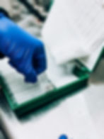
The merit of cell painting for SIOD researchers is the sheer volume of data. This standardized approach takes cells of interest and cultures them using 10 plates that each have 384 wells. Every well contains a couple thousand cells, and every cell is analyzed for 10,000 features.
Using automated analysis, Dr. Lewis and his team can sift and sort through the data, comparing the observable traits of SIOD with tens of thousands of other publicly available cell painting assays for different medical conditions—looking for clues. Other diseases that share similar phenotype signatures could lead the way to drug candidates for SIOD.
“As far as I can tell, we are pioneers in using these endothelial cells in cell painting assays,” says Dr. Lewis of the cells lining the blood vessels and lymphatic vessels. When creating the human cell model of SIOD, he and his team turned induced pluripotent stem cells into endothelial cells. This is allowing the researchers to examine how the condition impacts arteries, veins, and capillaries in kids with SIOD, given their elevated risk of stroke.
“This is potentially a way of turning the disease into a tool we could use to screen for drug therapies. We had to reduce the complexity of the disease in the living body to something that we think is representative of the disease mechanisms in tissue culture.” - Dr. Lewis
In February, the Lewis Lab will be applying for a Foundation Award for Discovery-Stage Research from the California Institute of Regenerative Medicine (CIRM). The team has been spending months preparing preliminary data and findings for the grant.
We’ve been throwing everything at it,” says Dr. Lewis. “The plan is to ask for at least two to three years of funding to get support for developing a drug that targets and prevents the strokes associated with SIOD. So, we’ve really pivoted for this philanthropy.”
What would the CIRM grant help to accelerate?
-
Basic science: Allowing researchers to better understand SIOD and verify results from lab models with real-life patient experiences and biology.
-
Drug screening: Letting scientists screen existing therapeutics very efficiently, up to 2,000 compounds at a time, in search of drugs that can benefit SIOD patients.
-
Clinical trials: Beginning trials with children who have SIOD, using the most promising drugs discovered.
Researchers in the Lewis Lab especially hope to compare proteins in blood samples from SIOD patients with the engineered endothelial cells, and anticipate seeing the same proteins altered in both places. While Dr. Lewis doesn’t yet know for sure what they will find comparing the human blood cells with the tissue culture designed to mimic the SMARCAL1 deficiency, he believes it could be a critical step toward validating their disease model.
“Our hope is that all of the abnormalities associated with SIOD reflect a common problem that may be correctable by a single drug therapy.”
David B. Lewis, MD
Thank you, Jessica and Kyle.
Because of your visionary leadership, and the contributions of everyone in the Kruzn for a Kure community, we are laying the groundwork for a better tomorrow for children with SIOD.
We will keep you informed of the results of the CIRM grant application. Thank you, as always, for everything you have made possible to bring us to this moment.
Michael Tomura
Associate Director, Major Gifts
June 15, 2022 Publication - New England Journal of Medicine Publication

Sequential Stem Cell–Kidney Transplantation in Schimke Immuno-osseous Dysplasia
Lifelong immunosuppression is required for allograft survival after kidney transplantation but may not ultimately prevent allograft loss resulting from chronic rejection. We developed an approach that attempts to abrogate immune rejection and the need for post-transplantation immunosuppression in three patients with Schimke immuno-osseous dysplasia who had both T-cell immunodeficiency and renal failure.
Each patient received sequential transplants of αβ T-cell–depleted and CD19 B-cell–depleted haploidentical hematopoietic stem cells and a kidney from the same donor. Full donor hematopoietic chimerism and functional ex vivo T-cell tolerance was achieved, and the patients continued to have normal renal function without immunosuppression at 22 to 34 months after kidney transplantation. (Funded by the Kruzn for a Kure Foundation.)